SONOANATOMY · BARCELONA
Anatomy made visible.
Discover the ultrasound and anatomical landmarks that bring clarity and confidence to musculoskeletal imaging. Sonoanatomy is a practical, multidisciplinary course designed to refine your orientation, localization and intervention skills across the spine and extremities.
19TH EDITION
Sono-Landmarks II
Small-group workshops, hands-on practice and direct interaction with expert tutors—supported by EULAR faculty and a multidisciplinary team of specialists.
22 & 23 January 2027
University of Barcelona · Bellvitge campus
THE COURSE
What is Sonoanatomy?
Sonoanatomy is an interactive and multidisciplinary course organized by Instituto Poal de Reumatología at the University of Barcelona, generally under the umbrella of EULAR and Euroson. It brings together applied clinical anatomy, ultrasound imaging and image-guided interventions in a highly practical learning environment.
Clinically relevant topics
Learn anatomy with direct relevance to daily clinical decisions, diagnosis and intervention.
Personalized training
Small groups and an interactive, multidisciplinary approach allow for individualized guidance.
Hands-on practicality
Move between anatomy and ultrasound through workshops designed around the level of participants, with time to practice and hone your skills.
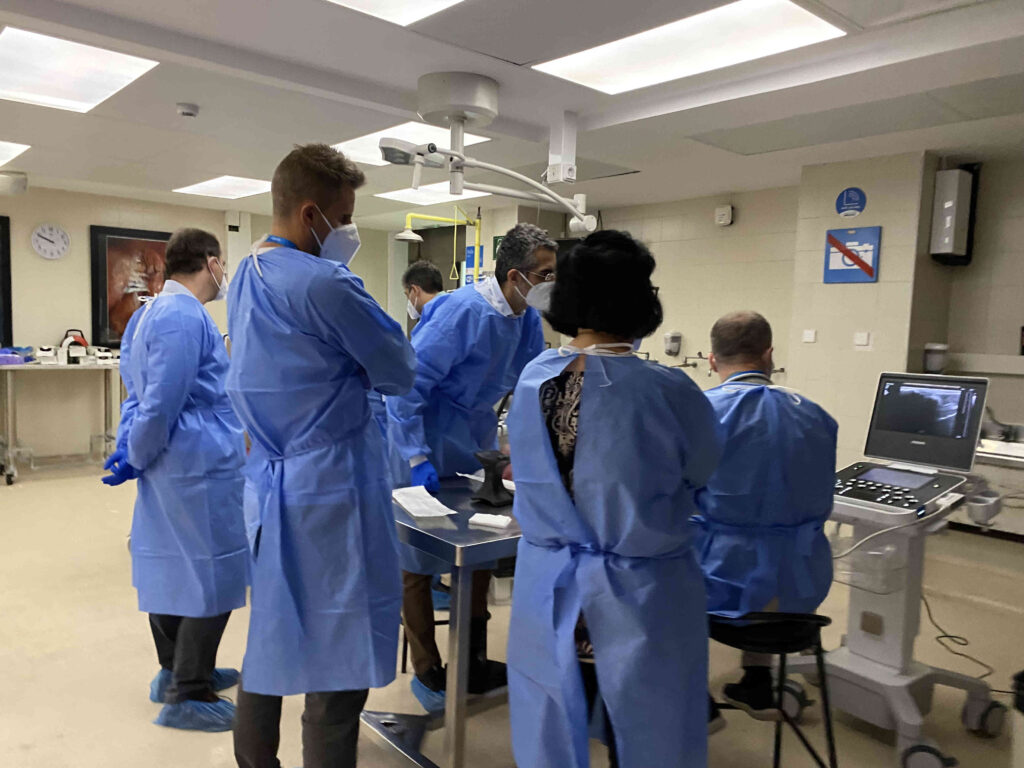
With more than 18 years of sonoanatomical and interventional training at the University of Barcelona, we avoid large auditorium lectures in favour of direct interaction, experienced faculty members and personalized guidance throughout the programme.
From applied clinical anatomy to ultrasound-guided procedures, the course gives healthcare professionals a comprehensive, practical journey that goes beyond the basics and builds confidence for day-to-day clinical practice.
THE 19TH EDITION
Sonoanatomy 2027
SONO-LANDMARKS II
The 19th edition of Sonoanatomy takes ultrasound anatomy beyond the obvious, exploring the axial body and extremities through advanced, landmark-based techniques.
Discover the ultrasound anatomy of fasciae, peripheral nerves, deep stabilizers, mini-entheses, retinacula, dynamic structures, and clinically hidden targets. Develop a systematic approach to both the axial musculoskeletal system and the extremities, integrating dynamic assessment and ultrasound-guided interventions.
Hands-on learning
The course places hands-on learning at its core, with intensive small-group workshops on cadaver specimens and live models. Participants move directly between anatomy and ultrasound, identifying landmarks, scanning structures in real time and practicing image-guided procedures under close faculty supervision.
An established learning environment
Built on more than 18 years of ultrasound education at the University of Barcelona, Sonoanatomy combines anatomical precision, clinical relevance and an exceptionally practical learning environment.
THE 19TH EDITION · SONOANATOMY 2027
Programme
Workshops utilizing cadaver specimens and models
Workshops from 8H30 until 18H – Lunch break 12H – 13H / 13H – 14H
Beyond the Spine
Friday, 22nd January 2027: 8H30 – 18H
Standardized landmark-based techniques for the ultrasound imaging of the axial musculoskeletal system, fasciae, abdominal wall, and clinically relevant peripheral nerves.
The spinal landmark map: muscles, ligaments, joints, and fascial planes
- Cervical, thoracic, and lumbar muscle compartments
- Posterior ligamentous complex
- Facet joint landmarks
- Interspinous and paraspinal anatomy
- Dynamic and functional anatomy of the spine
The abdominal wall and thoraco-lumbar fascia
- Rectus abdominis and linea alba
- External oblique, internal oblique, and transversus abdominis
- Thoraco-lumbar fascia and interfascial planes
- Quadratus lumborum and psoas landmarks
- Clinically relevant abdominal wall anatomy
Dynamic ultrasound of fasciae and interfascial planes
- Fascial gliding and anisotropy
- Functional anatomy of superficial and deep fasciae
- Ultrasound landmarks of interfascial compartments
- Fascial-related pain and edema patterns
Ultrasound landmarks of the peripheral nerves emerging from the spine
- Greater occipital nerve
- Dorsal scapular nerve
- Intercostal nerves
- Cluneal nerves
- Ilioinguinal and iliohypogastric nerves
- Lateral femoral cutaneous nerve
Ultrasound landmarks of axial entheses and deep stabilizers
- Multifidus and deep spinal stabilizers
- Enthesis-related anatomy of the axial skeleton
- Functional stabilizing structures of the trunk
Landmark-based ultrasound-guided interventions of the axial body
- Facet joint procedures
- Medial branch landmarks
- Interfascial injections
- Erector spinae and quadratus lumborum planes
- Hydrodissection techniques
Hidden Structures and Advanced Landmarks
Saturday, 23rd January 2027: 8H30 – 18H
Advanced landmark-based techniques for the ultrasound imaging of small structures, peripheral nerves, fasciae, dynamic stabilizers, and mini-enthesis organs.
The small entheses: pulleys, retinacula, and mini-enthesis organs
- Finger pulleys
- Extensor hood and sagittal bands
- Retinacula and small stabilizing structures
- Nail-enthesis unit
- Ultrasound anatomy of mini-entheses
Fascial and interfascial ultrasound landmarks of the extremities
- Fascia lata and iliotibial tract
- Brachial and antebrachial fasciae
- Fascial compartments of the extremities
- Dynamic fascial relationships
- Fascial-related pathology
The clinically hidden peripheral nerves
- Posterior interosseous nerve
- Superficial radial sensory nerve
- Dorsal ulnar sensory branch
- Baxter nerve
- Common fibular nerve
- Plantar interdigital nerves
Dynamic ultrasound of instability and motion
- Distal radio-ulnar joint dynamics
- ECU instability and subsheath anatomy
- Snapping elbow and snapping triceps
Snapping hip - Peroneal tendon instability
- Dynamic ankle stabilizers
The ultrasound landmarks of inflammation: synovio-entheseal and fascial functional units
- Functional enthesis concept
- Enthesis organ and fascia relationships
- Psoriatic arthritis-related landmarks
- Doppler assessment of small structures
- Ultrasound patterns of early inflammatory disease
Advanced landmark-based ultrasound-guided interventions of the extremities
- Tendon sheath procedures
- Small joint and retinacular interventions
- Peripheral nerve hydrodissection
- Interfascial procedures
- Ultrasound-guided procedures in small anatomical targets
FAQ for Attendees
Contacts for assistance
If you need any assistance these days, please contact the secretary team (Kirsten and Enrique)
- By e-mail: sonoanatomybcn@gmail.com
- By WhatsApp: +34 637542936 (Kirsten)
Sonoanatomy location
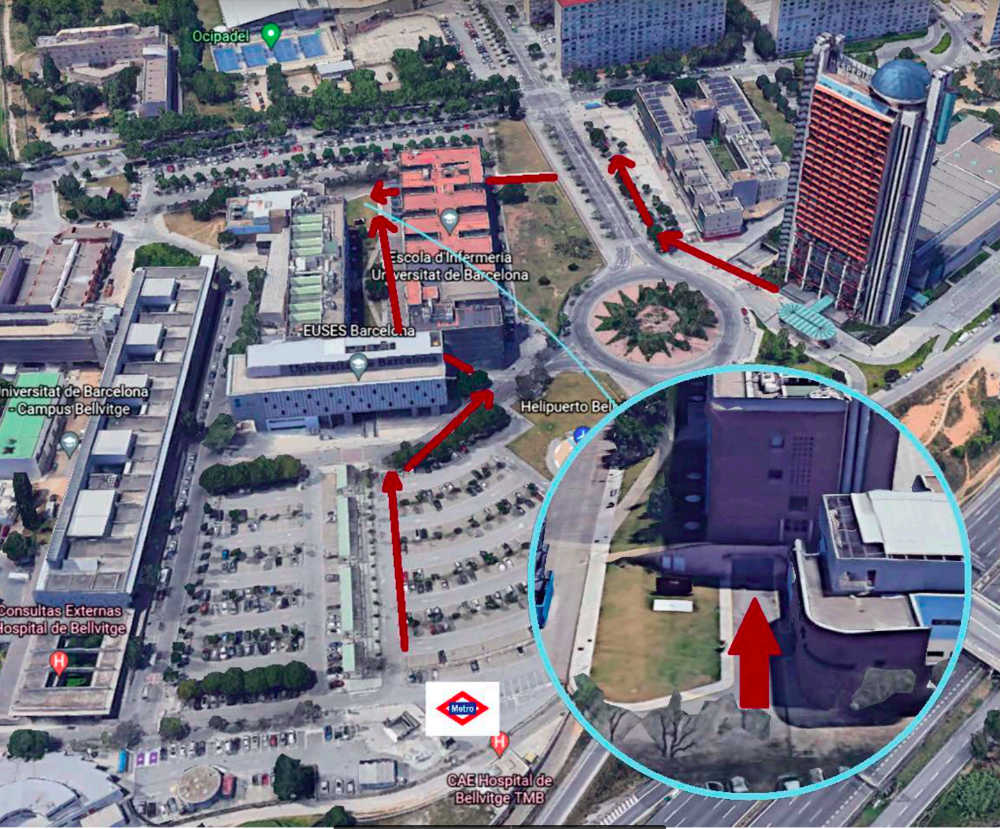
Sonoanatomy takes place in the dissection room of Universitat de Barcelona. This map will help you get to the Universitat de Barcelona:
How to get to the dissection room
Sonoanatomy takes place in the dissection room of Universitat De Barcelona. This image will help you understand how to get to the dissection room both from the Hotel Hesperia and from the nearest Metro (Hospital Belllvitge L1).:
Contact Us
If you prefer to contact us via email you can do it writing to sonoanatomybcn@gmail.com. If you are here you may be also interested on the personalized and tailored courses.